
What Does the Lump in This Image Appear to Be? A Detailed Explanation
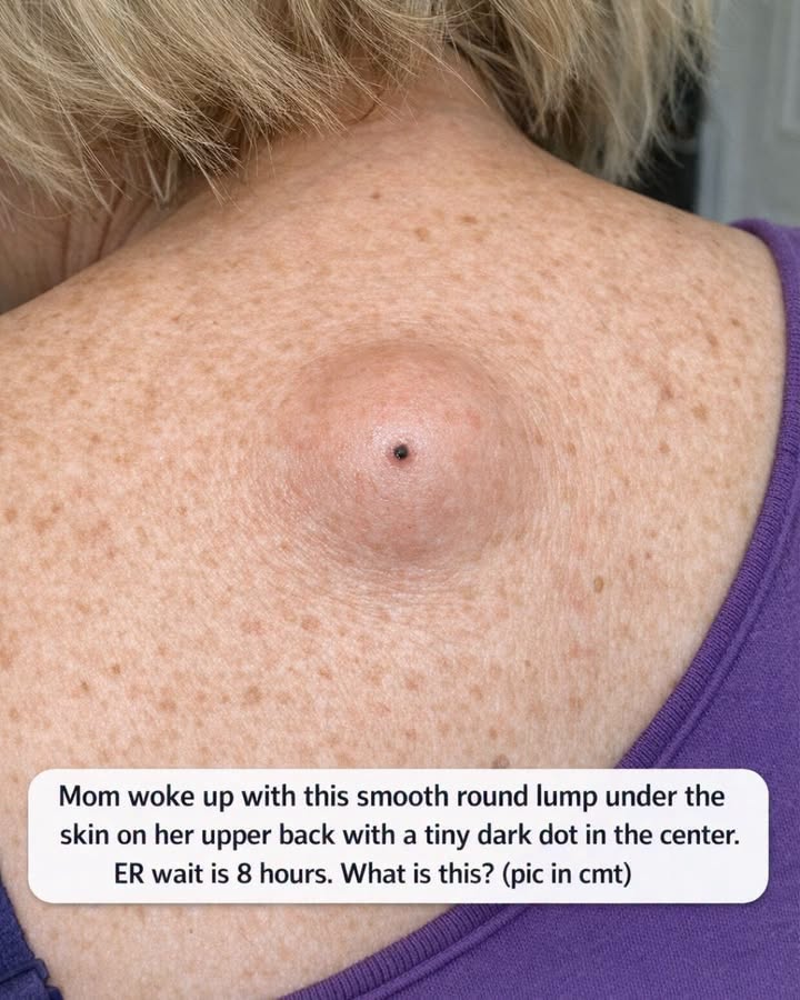
The photograph shows a smooth, rounded lump beneath the skin of the upper back, with a very small dark spot or “dot” in the center. Based on its visual appearance, the most likely explanation is an epidermoid cyst (also called an epidermal inclusion cyst). The combination of a dome-shaped lump and a central dark opening, known as a punctum, is particularly characteristic of this type of cyst.
However, a photograph alone cannot establish a medical diagnosis. A healthcare professional would normally examine the lump, feel whether it is mobile or fixed, assess its temperature and tenderness, and consider how quickly it developed.
What can be seen in the photograph?
The image shows the upper back and shoulder area of an adult person. Several features are visually important:
- There is a rounded, elevated swelling underneath the skin.
- The surface of the lump appears relatively smooth and intact.
- The swelling has a somewhat dome-like shape.
- A tiny black or dark central dot is visible.
- The surrounding skin does not appear to have extensive redness.
- There is no obvious open wound or large amount of drainage.
- The surrounding skin contains numerous small brown freckles or pigmentation spots.
- The lump appears to be located on the upper back, a common location for epidermoid cysts.
- The photograph does not contain a ruler or other reliable measurement reference, so its exact size cannot be determined from the image alone.
The central dark dot is the most diagnostically interesting feature. In an epidermoid cyst, this is called a punctum and represents an opening associated with the cyst. Medical references specifically describe a small, round lump with a central dark punctum as a typical appearance of an epidermoid cyst.
What is an epidermoid cyst?
An epidermoid cyst is a benign, usually non-cancerous sac beneath the skin. It contains a protein called keratin, along with dead skin cells and cellular debris.
Despite sometimes being called a “sebaceous cyst,” an epidermoid cyst is not actually a cyst of the sebaceous gland. “Sebaceous cyst” is a commonly used but technically inaccurate term for many of these lesions.
Epidermoid cysts most commonly occur on areas such as the face, neck and trunk, including the back. They can develop when skin cells that would normally shed from the surface become trapped deeper within the skin. Those cells continue producing keratin, gradually creating a sac filled with thick material.
Why is there a black dot in the middle?
The dark dot is called a punctum.
Think of the cyst as a small pocket beneath the skin. In some epidermoid cysts, the pocket has a tiny connection to the skin surface. That opening can become filled with keratin and appear as a black spot.
This is different from an ordinary blackhead. A blackhead is an open, clogged hair follicle, whereas the dark punctum associated with an epidermoid cyst is connected to an underlying cystic structure.
The punctum is one reason the lesion in this photograph looks more like an epidermoid cyst than a simple lipoma.
Why does the lump look so smooth?
Epidermoid cysts can sit beneath relatively normal-looking skin. When the cyst gradually enlarges, it pushes the surrounding skin outward, creating a rounded or dome-shaped elevation.
A cyst can therefore look surprisingly smooth and symmetrical, especially when it has not become inflamed.
The fact that the person in the photograph apparently noticed it upon waking does not necessarily mean the cyst formed overnight. It may have been developing unnoticed for a long time and only became noticeable because of its size, position, irritation, or a change in inflammation.
Epidermoid cysts are often slow-growing and painless.
Is it dangerous?
In most cases, an epidermoid cyst is not dangerous.
Many remain harmless for years and require no treatment if they are small, painless and not troublesome. However, cysts can sometimes become inflamed, infected, ruptured or increasingly painful.
Inflammation may cause the lump to become:
- Red
- Warm
- Tender
- Swollen
- Painful
- Larger than it previously was
If the cyst ruptures, thick yellowish or whitish material may come out. This material can have a strong or unpleasant odor because it contains keratin and cellular debris.
Could it be an abscess?
An abscess is another possibility whenever someone has a new lump, particularly if there is pain, redness, warmth or drainage.
However, the appearance shown here—with a smooth, rounded subcutaneous swelling and a distinct central punctum—is more suggestive of an epidermoid cyst than a typical skin abscess.
An abscess generally produces more obvious inflammatory changes, although an infected cyst can essentially behave like an abscess. A healthcare professional would distinguish between these conditions during examination.
Could it be a lipoma?
A lipoma is another common benign lump beneath the skin. Lipomas are made primarily of fatty tissue and frequently occur on the trunk and back.
However, lipomas generally do not have the characteristic central dark punctum seen in this image. The punctum therefore makes an epidermoid cyst a stronger visual possibility.
Other possibilities can include other types of skin cysts, inflamed follicles, benign tumors and, much less commonly, other skin lesions. The appearance alone cannot completely rule these out.
What would a doctor do?
A clinician can often recognize an epidermoid cyst through a physical examination. They may ask:
- When was the lump first noticed?
- Has it been growing?
- Is it painful?
- Is it warm or red?
- Has anything drained from it?
- Has it previously appeared and disappeared?
- Has the person had trauma or surgery in that area?
The clinician will also feel the lump to determine its consistency, mobility and attachment to deeper tissues.
Usually, additional imaging is unnecessary for a straightforward superficial cyst. In uncertain cases, an ultrasound can sometimes help characterize the lesion.
Does it need to be removed?
Not necessarily.
If an epidermoid cyst is small, painless and not causing problems, a doctor may recommend simply observing it.
If it repeatedly becomes inflamed, is painful, interferes with clothing, is cosmetically bothersome or continues to enlarge, removal may be considered.
A common definitive treatment is surgical excision, in which the cyst and its surrounding capsule are removed. Removing the entire capsule reduces the chance that the cyst will come back. Simply opening and draining a cyst can relieve pressure but may not eliminate the cyst because the capsule remains underneath the skin.
Should someone squeeze or puncture it?
No.
It is not advisable to squeeze, cut, puncture or attempt to drain a lump like this at home. Doing so can introduce bacteria, cause inflammation, create a wound and potentially make the lesion more difficult to treat. Cleveland Clinic specifically advises against trying to pop or drain an epidermal inclusion cyst yourself.
When would an emergency department be appropriate?
A stable, painless cyst without other symptoms generally does not automatically constitute an emergency. However, urgent medical assessment becomes more important if there is:
- Rapid enlargement
- Severe or rapidly increasing pain
- Significant redness or warmth
- Pus or foul-smelling drainage
- Fever or chills
- Red streaking spreading away from the lesion
- Significant bleeding
- The person becoming generally unwell
- A lump that is unusually hard, fixed or rapidly changing
Rapid growth or significant changes should be evaluated rather than assumed to be an ordinary cyst.
Overall interpretation of the image
The appearance is most consistent with an epidermoid (epidermal inclusion) cyst with a central punctum. The upper-back location, smooth rounded subcutaneous swelling and tiny dark central opening are a classic combination.
Importantly, this is an image-based impression, not a confirmed diagnosis. The photograph cannot determine whether the lesion is infected, how deep it extends, how long it has been present, or whether another type of lump is responsible.
If this photograph represents a real, newly discovered lump, particularly one that is growing, painful, red, hot or draining, it would be sensible to have it examined by a doctor or dermatologist. If it is painless and otherwise unchanged, it is usually reasonable to arrange a routine medical evaluation rather than attempting to remove it at home.
Bottom line: the little black dot is likely a punctum, and the overall appearance strongly resembles an epidermoid cyst—a usually benign cyst containing keratin beneath the skin.

